(15/25)
In honour of the Diefenbunker’s 25th anniversary, we continue to highlight 25 artifacts from within our collections. This week, with Halloween quickly approaching, we are illuminating all things skeletal and featuring an X-ray machine from the mid 1900s.
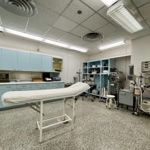 During the Diefenbunker’s operation as Canadian Forces Station (CFS) Carp between 1961 and 1994, the Medical Centre was equipped and supplied to be run by a doctor and nursing staff in the event of a nuclear attack. It contains a dispensary counter for pharmaceuticals, a lab area to perform tests, doctor’s office to examine patients, and a row of gurney and frame beds for the ill and injured. The Medical Centre had the capabilities to perform a variety of surgical procedures in a dedicated operating room, everything but open-heart and brain surgery, and in order to perform many of these operations, X-rays were required.
X-rays, also referred to as radiographs, were first observed and documented in 1895 by German physicist Wilhelm Conrad Röentgen. At the time of his discovery, Röentgen was investigating the effects of electron beams (rays) in electrical discharges through low-pressure gases and noticed that a fluorescent screen in his laboratory lit up when exposed to these invisible rays. Röentgen also discovered that detailed images were produced from firing streams of X-rays through arms and hands, outlining the bones within. Röentgen’s discovery of X-rays earned him the first Nobel Prize in Physics in 1901. To create a radiograph, the patient is positioned so that the part of the body being imaged is located between an X-ray source and an X-ray detector. When the machine is turned on, X-rays travel through the body and are absorbed in different amounts by different tissues, depending on the radiological density of the tissues they pass through. This electromagnetic radiation creates images of bones and other internal structures that are commonly used to diagnose bone fractures, infections, and other medical conditions.
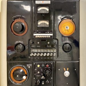 This X-ray power unit from the Diefenbunker’s collections was built by General Electric X-Ray Corporation between 1930 and 1951. This organization was founded by C.F. Samms and J.B. Wantz in 1893 where they originally specialized in electrostatic generators, however, General Electric X-Ray Corporation quickly delved into X-ray machine production in 1896, only one year after Röentgen’s discovery. Today, the corporation is known as GE Healthcare, and it continues to be a leading organization in manufacturing medical technology around the world.
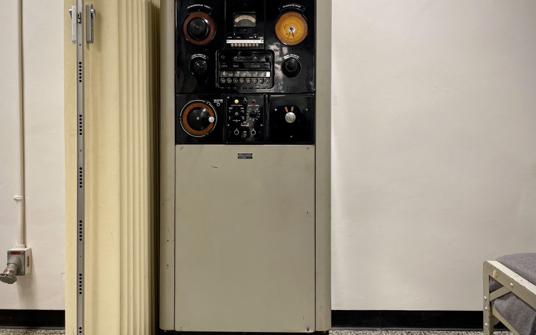
This X-ray machine stands 70” tall and is constructed with a metal frame that is tan in colour. There are several components on the face of the unit including silver and black metal dials, red and orange coloured plastic seen under both the Kilovolts Peak, a gauge used to monitor the maximum high voltage applied across an x-ray tube to produce the X–rays, and the Synchronous Timer reader, a timer that alternates energizing and de-energizing a load at a specific rate. Most of the dials and readers on the machine are meant to adjust and monitor the power flowing from the electrical unit through to the person or object being scanned.
Though it can no longer be used for radiograph imaging, this X-ray machine can still be viewed today in the Diefenbunker’s Medical Centre located on the 400 Level. Check it out on your next visit underground!
Stay tuned as we continue to celebrate our 25th anniversary by uncovering stories from our museum’s collections.